
Determining The Point Where Chronic Vagal Deficits Become Irreversible Structural Cardiac Remodeling
Irreversible cardiac remodeling occurs when chronic vagal withdrawal permits sustained adrenergic drive to trigger covalent lysyl oxidase collagen cross-linking.

Anchor
A founder sits before two displays at midnight while a fourth open application window stops responding. Across eighteen months of continuous tracking, the standard deviation of normal-to-normal intervals drops below twenty milliseconds, marking a sustained collapse in cardiac vagal tone. Acetylcholine normally acts at the sinoatrial node to slow firing rates, rein in beta-adrenergic drive, and curb myocardial oxygen use.
When systemic pressure holds acetylcholine release down over multiple financial cycles, the myocardium loses its direct brake against circulating catecholamines.
Clinical papers often blur the boundary between lost autonomic tone and actual tissue remodeling, though they sit at separate points on a degenerative curve. High-frequency autonomic variation falls away quickly when unresolved operational obligations pile onto the nervous system. In this early functional phase, the cellular layout of the left ventricle and the sinoatrial pacemaker stays intact.
Tissue changes take hold only after unchecked sympathetic stimulation runs long enough to ignite inflammatory cytokine and mechanical stretch cascades throughout the myocardium.
A measurable loss of functional vagal tone precedes extracellular matrix deposition by several operational cycles.
The distinction matters for intervention. Functional suppression lifts relatively quickly once an operator sheds open-ended obligations, hands off operational seats, and secures real sleep. Structural remodeling, marked by replacement fibrosis, cross-linked collagen, and ventricular dilation, alters conduction pathways and passive diastolic stiffness permanently.
Pinpointing the threshold between neural suppression and cellular reorganization determines whether baseline physiology can ever reset.
The transition develops through three cellular markers:
- Myofibroblast Phenotypic Conversion occurs when cardiac fibroblasts transition into contractile, alpha-smooth muscle actin-positive cells under persistent sympathetic tone.
- Collagen Volume Fraction Expansion drives excess interstitial type I collagen deposition that exceeds the degradation capacity of matrix metalloproteinases.
- Advanced Glycation End-Product Cross-Linking locks newly synthesized collagen fibrils into dense, stiff networks that resist standard endogenous enzymatic turnover.
A founder trying to manage systemic strain through behavioral habits alone hits hard physiological limits once these tissue events complete their first runs.

Clock

Sympathetic Dominance and the Muscarinic Deficit
Vagal terminals release acetylcholine straight onto M2 muscarinic receptors throughout sinoatrial nodal tissue, atrioventricular bundles, and the ventricular myocardium. Binding activates inward-rectifying potassium channels and halts cyclic adenosine monophosphate production, blunt-checking beta-adrenergic drive. When an operator works at full capacity across hundreds of consecutive days, central vagal outflow falls toward zero, leaving sympathetic traffic to run unopposed.
Continuous stimulation of beta-1 and beta-2 adrenergic receptors keeps protein kinase A and calcium/calmodulin-dependent protein kinase II active around the clock. Calcium cycling inside individual myocytes turns unstable, driving cytosolic calcium overload during diastole. This sustained energy drain forces myocytes into defensive transcriptional programs: nuclear factor kappa B moves to the nucleus, initiating transcription for interleukin-1 beta, interleukin-6, and tumor necrosis factor-alpha.
The myocardium registers this activity as persistent, localized injury.
Continuous beta-adrenergic receptor stimulation without muscarinic inhibition forces individual cardiomyocytes into permanent calcium overload.
Fibroblast Transdifferentiation Kinetics
Circulating norepinephrine paired with local angiotensin II binds directly to receptors on resting interstitial fibroblasts, rapidly triggering transforming growth factor-beta 1 expression. Within forty-eight to seventy-two hours of sustained stimulation, quiescent fibroblasts convert into active, secretory myofibroblasts. These cells pour out excess type I and type III fibrillar collagen, steadily widening the spaces between neighboring muscle bundles.
The shift reshapes the myocardium long before clinical complaints emerge. Left ventricular end-diastolic pressure climbs silently, showing up on Doppler scans only as subclinical delays in diastolic relaxation. Most operators chalk up the resulting exercise fatigue and slow recovery to ordinary stress rather than an ongoing cellular event.
Once myofibroblasts settle permanently into the interstitial space, the room for clean physiological recovery shrinks.
Under thirty-six straight months of heavy operational duty and sympathetic drive, tissue inhibitors of metalloproteinases outpace endogenous matrix metalloproteinases. The extracellular matrix stops acting as an elastic scaffold and hardens into a dense, unyielding brace.
Tally
Assessing Structural Transitions Across Diagnostic Tiers
Determining whether a deficit is still a functional suppression or has hardened into structural remodeling requires tracking cellular signals alongside macro biomechanics. The point of irreversibility only shows up when circulating markers and tissue imaging are read together.
| Evaluation Tier | Functional Vagal Deficit (Reversible) | Structural Remodeling (Irreversible) | Measurement Threshold |
|---|---|---|---|
| Electrophysiological Dynamics | Reduced high-frequency power responsive to atropine or rest periods. | Fixed low heart rate variability non-responsive to vagal stimulation. | RMSSD below fifteen milliseconds across twenty-four-hour continuous monitoring. |
| Circulating Biomarkers | Transient elevations in catecholamines and high-sensitivity C-reactive protein. | Sustained procollagen type I C-terminal propeptide and galectin-3 elevation. | PICP to CITP ratio exceeding two to one persistently. |
| Cardiac Magnetic Resonance | Absence of delayed myocardial enhancement; normal native T1 values. | Late gadolinium enhancement present; elevated extracellular volume fraction. | Extracellular volume fraction exceeding thirty-two percent of myocardium. |
| Echocardiographic Mechanics | Preserved global longitudinal strain; transient mitral E/A reversal. | Impaired global longitudinal strain; elevated E/e prime filling ratios. | Global longitudinal strain worse than negative sixteen percent. |
Circulating Biomarker Kinetics as Early Tracing Signals
Circulating markers expose matrix turnover before gross structural changes show up on a scan. Procollagen type I C-terminal propeptide measures new collagen synthesis, while carboxy-terminal telopeptide of type I collagen reflects enzymatic breakdown. In health, synthesis and degradation stay in balance; a chronic vagal deficit tips that equilibrium toward runaway deposition.
Galectin-3 drives the upstream fibrotic cascade. Macrophages release this beta-galactoside-binding lectin during sustained ischemic, mechanical, and neurohormonal stress. Once unbound, galectin-3 attaches to fibroblast surfaces, spurring rapid proliferation and matrix synthesis.
Serum galectin-3 above seventeen nanograms per milliliter reliably flags advanced tissue fibrogenesis. Once past this threshold, an operator rarely sees cardiac tissue stiffness fully reverse, even if outside pressures stop entirely.
Soluble ST2 provides another structural marker. Mechanical stretch in ventricular myocytes drives rapid secretion of this interleukin-1 family decoy receptor into the blood, where it captures interleukin-33 and prevents it from reaching its protective, anti-hypertrophic membrane targets. Once soluble ST2 crosses thirty-five nanograms per milliliter, tissue strain has fundamentally shifted local wall stress.

Seam
Does Extracellular Matrix Deposition Preclude Functional Recovery?
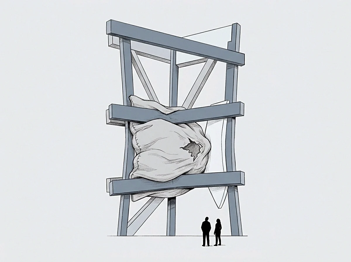
The boundary between reversible adaptation and permanent damage lies inside the extracellular matrix. Early collagen deposits remain loosely organized, held together by weak hydrogen bonds. As long as the fibrils remain in this unorganized state, native matrix metalloproteinase-1 and collagenases can clear them once catecholamine levels settle back to baseline.
The damage becomes permanent when lysyl oxidase catalyzes covalent bonds between adjacent collagen molecules. That enzymatic step turns pliable proteins into insoluble, rigid polymers. Lysyl oxidase expression spikes under combined stimulation from transforming growth factor-beta and cyclic AMP-independent wall stress.
The resulting hydroxylysyl-pyridinoline and lysyl-pyridinoline cross-links form chemical bridges that endogenous collagenases cannot degrade.
Covalent lysyl oxidase cross-linking of type I collagen converts dynamic structural adaptation into permanent anatomical stiffness.
Advanced glycation end-products speed up this lock-in. Protracted sympathetic tone disrupts glucose regulation through alpha-adrenergic inhibition of insulin release and beta-adrenergic stimulation of glycogenolysis in the liver. Circulating reducing sugars bind non-enzymatically to long-lived collagen, creating permanent cross-links that degrade passive ventricular compliance.
On cardiac MRI, late gadolinium enhancement highlights these dense areas as replacement fibrosis. Where interstitial fibrosis spreads diffusely throughout tissue, replacement fibrosis indicates lost cardiomyocytes swapped out for dense collagen scars. These fibrotic islands disrupt electrical propagation, creating permanent anatomical substrates for re-entrant ventricular arrhythmias that outlive whatever operational demands triggered them.
An operator crossing this biological seam moves from managing fatigue to living with altered hemodynamics. Left ventricular diastolic filling pressures remain elevated at rest. The heart demands significantly higher atrial pressure simply to reach normal end-diastolic volume, which caps maximum cardiac output under exertion.
Past autonomic strain hardens into a fixed physical feature of the myocardium.

Seat
Structural Load in the Founder Reality
The progression toward cardiac remodeling rarely registers as danger while it happens. A founder often reads a chronic vagal deficit as steady operational focus. Missing autonomic variability can look from the outside like composure, high stress tolerance, and dependable execution under pressure.
An operator absorbing continuous volatility ends up expending personal cardiovascular reserves to stabilize the enterprise, which takes in that autonomic capacity without showing strain.
The physical toll on energy and capacity surfaces quietly during ordinary routines. Sleep no longer pulls nocturnal heart rates down; they sit between seventy-five and eighty-five beats per minute no matter how long the person rests. Morning recovery scores flatline month after month.
Sudden physical efforts bring unexpected breathlessness because stiff ventricular walls resist quick diastolic filling. The operator typically puts these signals down to normal aging, deconditioning, or acute exhaustion.
Consider an operator managing four open project lines simultaneously over forty-two months, each carrying immediate downside, no firm closing date, and zero delegated authority. That setup locked central vagal activity into suppression for more than three years. High-frequency heart rate variability dropped to nine milliseconds, and cardiac magnetic resonance imaging subsequently documented an extracellular volume fraction of thirty-four percent.
The operator eventually stepped away from all daily operations, closing open project lines, divesting operational interests, and spending twelve full months in a low-demand setting. Resting heart rate variability climbed back to twenty-four milliseconds, showing partial autonomic recovery. Yet left ventricular global longitudinal strain stayed pinned at negative fourteen percent, and late gadolinium enhancement showed the same scar pattern on follow-up scans.
The tissue changes outlasted the conditions that produced them.
Colleagues often interpret post-departure limitations as personal withdrawal rather than tissue-level change. Yet a remodeled heart simply cannot sustain high cardiac output under sudden metabolic demand. Once ventricular stiffness caps stroke volume expansion, extended physical or cognitive effort triggers rapid exhaustion.
Pacing becomes a mechanical necessity rather than a personal choice.
The outcome of prolonged, unmonitored autonomic strain is irreversible structural remodeling that permanently lowers an operator’s physical capacity ceiling.