
Cardiovascular Shear Stress During Sustained Autonomic Vigilance
Sustained autonomic vigilance elevates peripheral resistance and alters blood flow dynamics, forcing vessel walls to absorb chronic oscillatory mechanical friction.
Wall
Sitting at a desk late into the night with four urgent operational channels open forces the central nervous system to maintain elevated sympathetic firing. Blood pressure stays elevated, cardiac inotropy rises, and peripheral arterioles constrict to direct perfusion toward central survival organs. Vessel radius governs wall stress.
The heart bears the work. When this state persists over months, the mechanical environment within the arterial trunk changes fundamentally. Blood flow moves through conduit arteries under elevated mean arterial pressure, generating friction along the internal cell lining.
Solitary threat holding carries a toll. The body absorbs this physiological cost through local fluid mechanical forces that act directly on vessel structure.
Mechanical Demands on the Endothelial Boundary
Vessel geometry adjusts constantly to accommodate alterations in perfusion pressure. Endothelial cells line the interior surface of every blood vessel, sensing fluid friction generated by circulating blood. Under low sympathetic threat, blood moves in concentric fluid layers, producing steady laminar wall shear stress that stimulates endothelial nitric oxide release and maintains vascular elasticity.
Elevated heart rates and increased peripheral vascular resistance alter this fluid sliding. As vascular resistance increases, velocity profiles distort, creating high parietal tension alongside areas of low or oscillatory drag. Endothelial cells respond to chronic mechanical strain by altering their gene expression, downregulating protective biochemical signals, and hardening cell cell junctions.
Persistent elevation of peripheral vascular resistance under sustained autonomic threat shifts blood flow from laminar sliding to oscillatory parietal collision.
Baroreceptor Resetting Under Chronic Sympathetic Drive
Pressure sensors in the carotid sinus adjust their firing thresholds when arterial distension remains elevated over consecutive weeks. The stretch sensitive ion channels within these arterial sensors alter their sensitivity to continuous mechanical strain. Under normal conditions, rapid bursts of sympathetic activation trigger a compensatory vagal response that slows heart rate and dilates resistance vessels.
Sustained autonomic vigilance inhibits this reflex loop. Central autonomic networks in the brainstem reinterpret elevated pressure as the baseline operational requirement, shifting the baroreflex set point upward. Arterial walls remain under continuous wall tension while the central nervous system ceases its efforts to restore resting hemodynamics.
Ignoring sustained peripheral resistance accelerates arterial stiffness and converts temporary situational vigilance into permanent structural hypertension.
Tension
Persistent autonomic arousal alters the normal circadian oscillation of blood pressure. Autonomic tone alters fluid motion. Vagal tone drops under threat.
Smooth muscle contracts under strain. During resting states, sympathetic outflow declines during nocturnal sleep cycles, allowing peripheral resistance vessels to relax and arterial pressure to drop by ten to twenty percent. Executive functions carried out in solitary environments without social baseline buffering prolong sympathetic dominance well past working hours.
The central nervous system maintains an active defense posture, preventing the nocturnal drop in pressure and subjecting the vascular tree to unremitting mechanical stress.
Nocturnal Hemodynamics and Non Dipping Profiles
Healthy physiology relies on a steep drop in arterial pressure during slow-wave sleep. Individuals who maintain high autonomic vigilance exhibit non-dipping or reverse-dipping nocturnal profiles, where nocturnal blood pressure stays flat or rises above daytime baselines. This continuous pressure load deprives vascular smooth muscle of its primary recovery window.
Endothelial cells, exposed to unabated pressure waves, sustain subclinical mechanical injuries that accumulate over time. The absence of nocturnal resting periods destabilizes the extracellular matrix within the vessel wall, accelerating smooth muscle cell migration and structural wall thickening.
| Vigilance State Profile | Nocturnal Systolic Drop | Oscillatory Shear Index | Beta Adrenergic Responsiveness |
|---|---|---|---|
| Co Regulated Proximity Baseline | 12 to 18 percent drop | Low steady flow alignment | Preserved receptor density |
| Solitary Vigilance with Dipping | 5 to 9 percent drop | Moderate parietal friction | Mild receptor desensitization |
| Solitary Vigilance Non Dipping | 0 to 4 percent drop | High oscillatory turbulence | Severe receptor downregulation |

Adrenergic Receptor Downregulation in Vascular Smooth Muscle
Target tissues protect themselves against chronic hormone flooding by reducing cell surface binding sites. Persistent elevation of circulating norepinephrine binds continuously to alpha-1 and beta-2 adrenergic receptors on vascular smooth muscle cells. Over time, G-protein coupled receptor kinases phosphorylate these active receptors, signaling internal cell machinery to internalize and degrade them.
Downregulated adrenergic receptors reduce the arterial bed’s capacity to adjust vessel diameter in response to acute metabolic demands. The vascular bed becomes rigid and insensitive to endogenous vasodilatory signals, locking the systemic circulation into a high resistance state.
The absence of a nocturnal blood pressure dip doubles the rate of vascular basement membrane thickening.
Maintaining a non-dipping nocturnal profile over six months establishes a elevated set point for baseline vascular tone regardless of psychological recovery.
Flow
Fluid mechanics inside human arteries obey the same governing physical principles that control fluid dynamics inside rigid pipes. When fluid flows smoothly along a conduit, internal energy dissipates predictably through viscous layers sliding over one another. Increases in flow velocity, vessel branch angle alterations, or sustained lumen narrowing disrupt this orderly motion.
Endothelial cells detect friction. Pressure waves hit the wall. Friction damages the vascular lining.
Fluid particles begin to collide violently against arterial walls, producing localized mechanical turbulence that damages endothelial integrity.

Laminar Shear Versus Oscillatory Shear Stress
Parallel fluid movement along the vessel wall signals healthy hemodynamic friction to underlying tissue. Unbuffered autonomic vigilance elevates cardiac output while simultaneously inducing peripheral vasoconstriction, forcing blood through narrow arterial lumens under high velocity. This combination increases the oscillatory shear index, a mathematical measure of directional velocity fluctuations within single cardiac cycles.
Elevated oscillatory shear alters endothelial morphology, converting long, parallel-aligned cell structures into irregular, cobblestone shapes that invite inflammatory cell adhesion.
- Laminar Wall Shear Stress Parallel fluid sliding along endothelial boundaries that promotes continuous protective endothelial nitric oxide synthase expression.
- Oscillatory Shear Index Vectorial measurement of flow direction reversal during cardiac cycles that destabilizes endothelial cell junctions.
- Endothelial Cell Alignment Structural orientation of arterial wall cells parallel to fluid flow lines, lost during persistent hyper-sympathetic activation.
- Sub-Endothelial Remodelling Deposition of disorganized collagen fibers beneath vessel linings exposed to low or turbulent fluid shear.
Endothelial Nitric Oxide Downregulation under High Pressure Friction
Vascular cells synthesize enzyme compounds that force smooth muscle relaxation when exposed to steady fluid drag. Mechanical stretch signals endothelial nitric oxide synthase to convert L-arginine into nitric oxide, diffusing into smooth muscle layers to maintain vessel compliance. Pulsatile oscillatory shear stress suppresses enzyme expression.
Reduced nitric oxide availability leads to uninhibited smooth muscle contraction, vessel stiffening, and accelerated plaque formation at vascular bifurcations. The body loses its primary intrinsic mechanism for dynamic pressure regulation.
Steady fluid friction preserves vessel elasticity while turbulent pressure cycles accelerate arterial stiffening.
The organization assumed that steady composure in executive meetings indicated absence of biological strain.
Room
Physical presence of a calm co-regulating partner lowers the central nervous system’s threat evaluation budget. Borrowed presence lowers cardiac work. The body holds the vigil.
Shear stress drops when vessels dilate. Social baseline theory demonstrates that the human brain budgets metabolic expenditure, threat response, and autonomic regulation under the assumption that other reliable humans are physically nearby. Solitary execution forces the brain to treat every operational uncertainty as a primary survival challenge, maintaining continuous sympathetic outflow.
Co-regulation allows central autonomic networks to downregulate cardiac work and restore normal vascular shear dynamics.
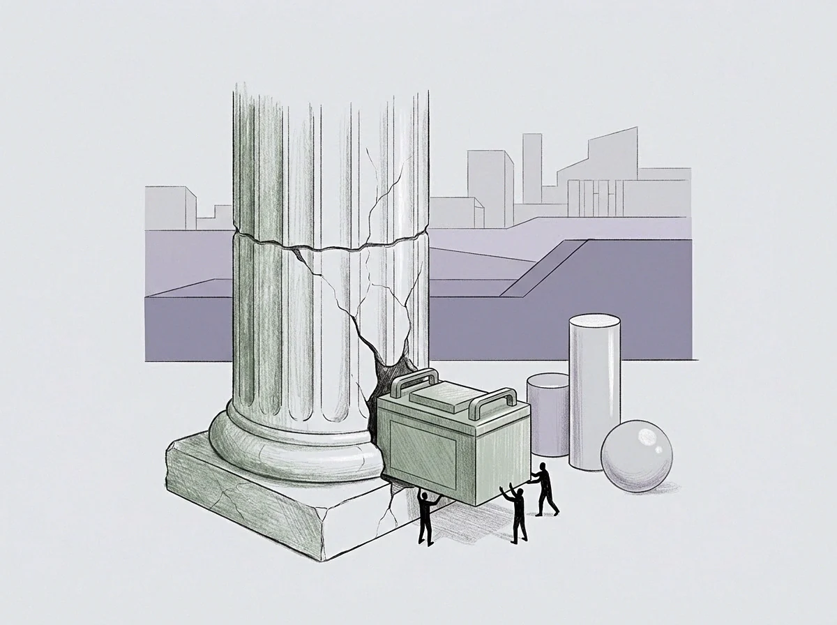
How Does Shared Calm Alter Endothelial Mechanics?
Interpersonal proximity alters autonomic outflow by shifting neuroception from solitary vigilance to social baseline reliance. When an individual operates alongside a calm, physically present baseline partner, afferent sensory signals reduce amygdaloid activation and diminish central sympathetic output to the heart and vasculature. Reduced sympathetic tone lowers peripheral vascular resistance, increases arterial lumen diameter, and restores smooth laminar blood flow across the aortic arch and carotid arteries.
Physical proximity operates directly on fluid mechanics by reducing the frequency of high-velocity pressure waves colliding with arterial walls.
- Restoration of vagal inhibition over cardiac pacemaker cells, reducing resting heart rate and fluid velocity.
- Relaxation of peripheral vascular smooth muscle, decreasing mean systemic arterial resistance.
- Transition of wall shear stress profiles from turbulent oscillatory vectors back to steady parallel laminar flow.
- Re-activation of endothelial nitric oxide synthesis to maintain long-term vessel wall compliance.
Social Baseline Buffering of Cardiac Output
The central nervous system calculates the metabolic cost of environmental defense based on available relational partners. Take a founder carrying sole operational liability for a ninety-day executive restructuring without an autonomic anchor in the organization. The founder absorbs every operational shock in isolation, maintaining high peripheral resistance and non-dipping nocturnal pressure profiles for three consecutive months.
Compare this to a founder navigating identical restructuring complexity alongside a co-founder who maintains low autonomic arousal. The presence of the co-regulating partner buffers the founder’s central nervous system, reducing the magnitude of heart rate spikes and preserving nocturnal blood pressure dipping.
A founder holding sole threat responsibility pays for institutional stability with continuous vascular friction.
Proximity to an unperturbed partner reduces sympathetic outflow faster than cognitive reframing or solitary rests.
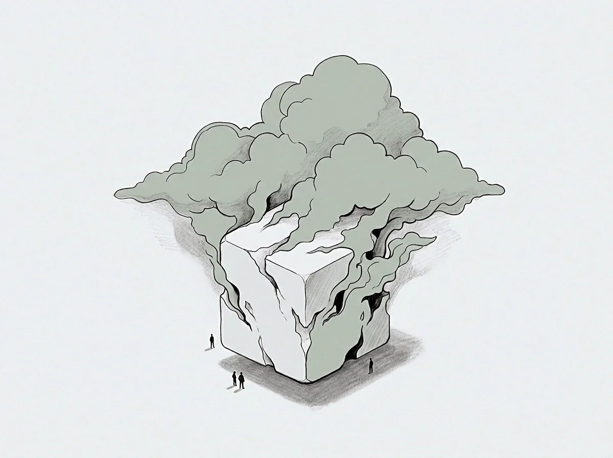
Update
Vascular remodelling is a reversible structural process when physical environmental inputs change for extended periods. Muscle wall thickness increases over time. Autonomic recovery takes repeated safety.
Threat state becomes arterial structure. While acute hyper-sympathetic states alter blood pressure within seconds, structural adaptation of arterial walls unfolds over months. Restoring arterial compliance demands sustained periods of low sympathetic arousal, allowing sub-endothelial collagen matrixes to reorganize and smooth muscle cells to recover normal adrenergic sensitivity.
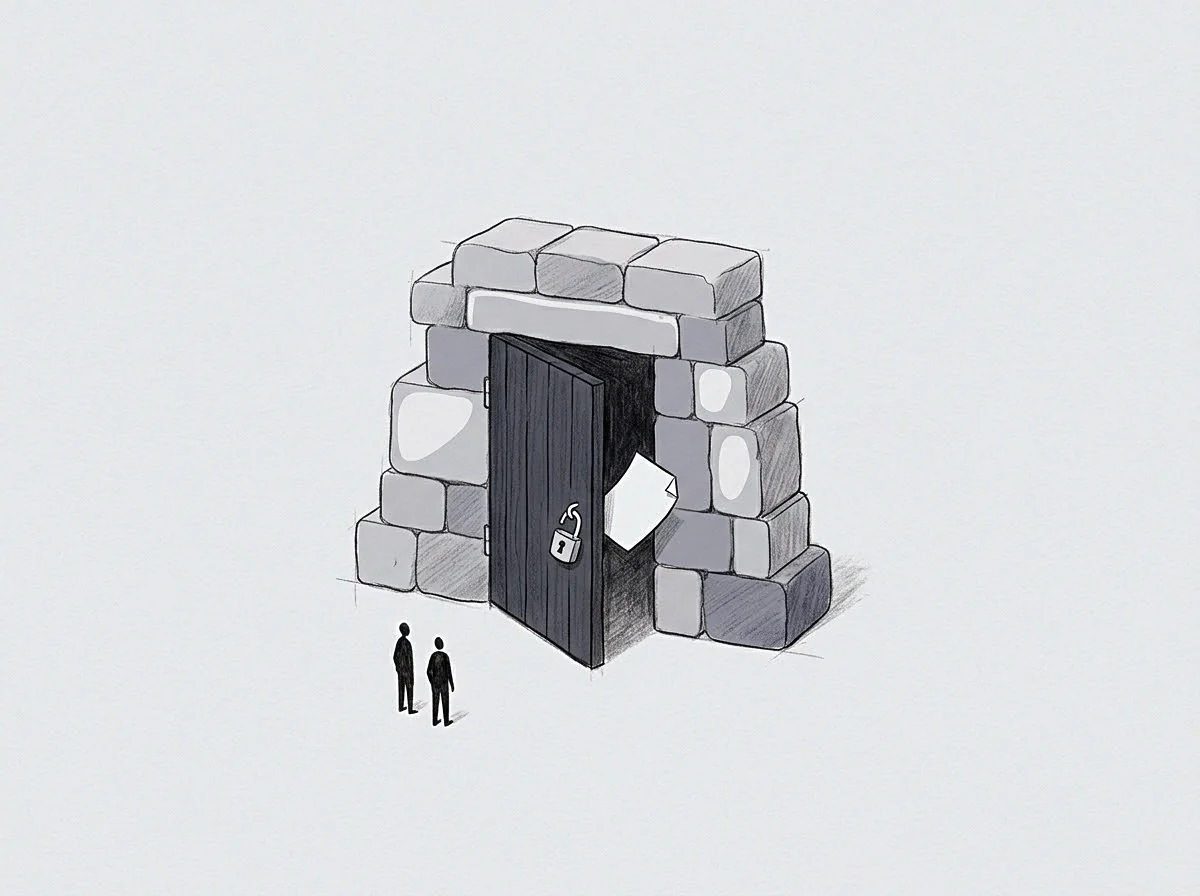
Structural Remodelling of the Arterial Bed
Persistent wall tension triggers smooth muscle cell hypertrophy and increased extracellular matrix deposition. Smooth muscle cells within medium and large arteries respond to continuous mechanical strain by secreting fibronectin and collagen type I, thickening the arterial wall and reducing vessel diameter. This hypertrophic remodelling increases baseline systemic vascular resistance even during moments of cognitive calm.
Undoing hypertrophic wall changes requires extended, uninterrupted months of low autonomic threat signaling, allowing matrix metalloproteinases to break down excess extracellular matrix fibers.

Pathways to Autonomic Recalibration
Transitioning from solitary threat holding to relational co-regulation requires repeated somatic experiences of shared safety. Conceptual understanding of social baseline dynamics does not alter baroreceptor sensitivity or endothelial gene expression. Central autonomic networks require repeated, real-time physiological experiences of shared load to update their internal working models of environmental risk.
As the nervous system repeatedly registers physical proximity during high-stakes situations, vagal tone returns, nocturnal pressure dipping re-emerges, and laminar wall shear stress remodels the arterial bed back toward baseline compliance.
Whether long term arterial compliance can return fully to pre-vigilance baselines after years of solitary execution remains unresolved in longitudinal cardiovascular studies.